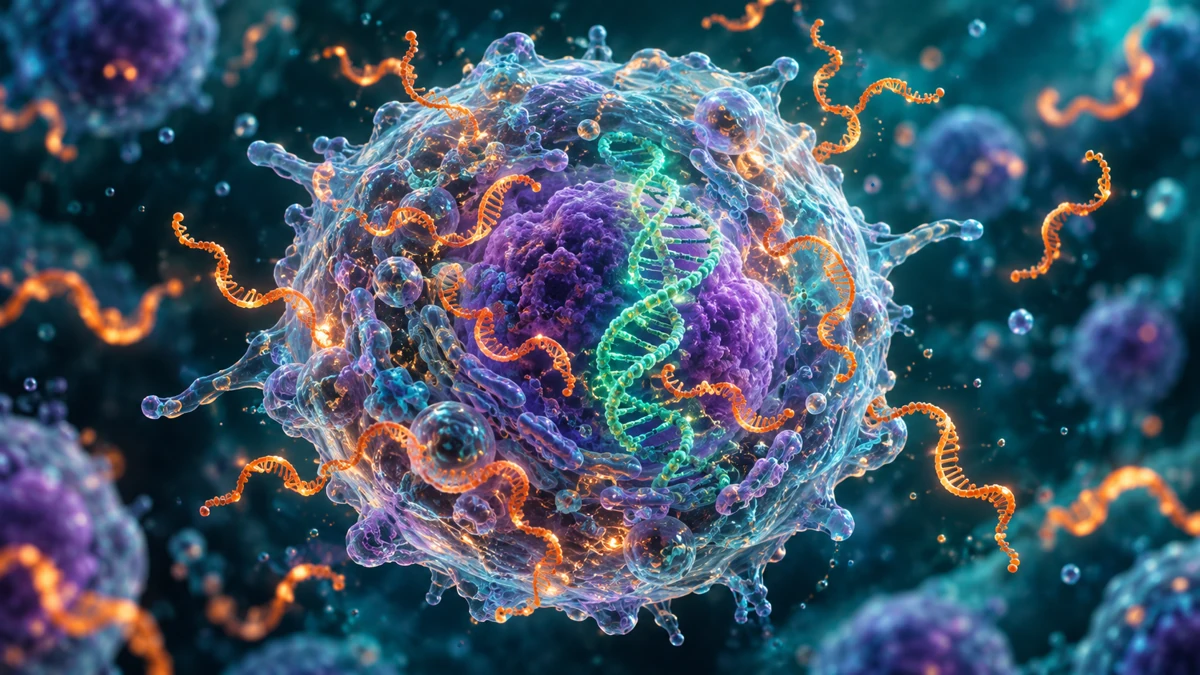
DNA reveals genomic alterations. RNA adds information about how genes are expressed and how their messages are joined. Together, these perspectives help build a more complete understanding of the disease.
⤨
DNA
Mutations
Copy number changes
Chromosomal abnormalities
+
∿
RNA
Gene fusions
Gene expression
Splice variants and clonality
Complementary insights. One comprehensive profile.
DNA: Define the Genomic Changes
DNA sequencing evaluates mutations and chromosomal changes that help characterize hematologic disease. These findings gain meaning when interpreted with blood counts, bone marrow morphology, immunophenotyping and the patient’s history.
A mutation does not answer every diagnostic question on its own. Integrating several types of molecular evidence helps clinicians assess whether the overall pattern fits the suspected disease.
RNA: Add Expression, Fusions and Clonality
RNA profiling evaluates gene messages, including expressed fusions and patterns of gene expression. It can add information about disease identity and abnormal splicing, while B- and T-cell clonality analysis contributes another view of lymphoid populations.
These complementary findings help clinicians interpret the molecular profile in a disease-specific context rather than relying on an isolated alteration.
Bring the Findings Together
Hematology Profile Plus combines mutation, fusion and expression analysis. The goal is an integrated evaluation that supports the clinical question—whether that involves leukemia, lymphoma, myeloma or another hematologic neoplasm.
For clinicians
Insights That Support Clinical Decisions
Understand the findings in the context of the patient’s diagnosis, pathology and treatment history.
01 / UNDERSTAND
Disease Characterization
Integrate mutations, fusions and gene expression with the clinical and pathology evaluation.
02 / EVALUATE
Treatment Biomarkers
Assess findings that may help inform discussions of targeted therapies, immunotherapies or clinical trials.
03 / INTEGRATE
Diagnostic Support
Use molecular findings to support classification and interpretation within the broader diagnostic workup.
Support Disease Classification
Mutations, fusions, chromosomal changes and expression patterns can contribute to disease classification. In lymphoma, expression-based information can support cell-of-origin assessment. Molecular findings should be interpreted alongside morphology, immunophenotyping and other diagnostic evidence.
Inform Prognostic Evaluation
Risk assessment draws on both disease biology and the patient’s clinical features. An integrated molecular profile gives clinicians additional findings to consider when evaluating prognosis and discussing the next steps in care.
Evaluate Treatment-Relevant Findings
Clinicians can review relevant molecular findings when considering treatment options or clinical trials. The significance of a finding depends on the specific diagnosis, treatment setting and available evidence; detecting an alteration does not guarantee a treatment match.
Understand the Broader Marrow
In multiple myeloma, evaluating CD138+ plasma cells and the CD138− population provides distinct perspectives. Plasma-cell profiling informs disease characterization, while evaluation of the remaining cell population can help assess possible co-existing myeloid disease.
Myeloma evaluation
CD138+ Plasma CellsCharacterize the plasma-cell disease
+
CD138− Cell PopulationAssess possible co-existing myeloid disease
One bone marrow. Complementary perspectives.
Conceptual workflow for myeloma evaluations. Relative cell proportions are not represented.
Additional capabilitiesB- & T-cell clonalityIGHV mutation statusHLA class IEBV
The relevance of each finding depends on the disease and specimen. Interpret results with the complete clinical workup.
Across hematology
One Profile. Multiple Clinical Applications.
A shared molecular approach, with interpretation guided by the clinical question.
Leukemia & Myeloid Neoplasms
Mutation and fusion analysis for AML, ALL and other hematologic neoplasms.
Lymphoma & Cutaneous Lesions
Expression and clonality insights, including DLBCL cell-of-origin assessment. Skin biopsies are accepted.
Multiple Myeloma
CD138+ plasma-cell profiling and CD138− evaluation to assess possible co-existing myeloid disease.
A Broader View of the Bone Marrow
For myeloma evaluations, looking at both cell populations adds context beyond the plasma-cell compartment.
♡
For patients & families
Understanding Your Blood Cancer’s Molecular Profile
This test examines DNA and RNA in a sample selected by your care team. It may help your doctor understand the molecular features of your blood cancer and discuss what they mean for your care.
Why has my doctor ordered this test?
To learn more about your disease’s molecular characteristics and how those findings may relate to your care.
What type of sample will I need?
Depending on your condition, your doctor may select blood, bone marrow or tissue. Your care team will explain the collection process.
Will this identify a treatment?
Results may inform options, but a treatment match is not guaranteed. Your doctor will explain what the findings mean for you.
Hematology Profile Plus combines DNA analysis with RNA profiling. RNA adds information about fusions, gene expression, abnormal splicing and clonality that complements DNA findings.
What type of sample does the test use?
Accepted specimens include bone marrow, peripheral blood, fresh tissue, skin biopsies and FFPE material. The appropriate sample depends on the condition being evaluated. Review the specimen requirements before collection.
How long do results take?
GTC’s current test page lists a turnaround time of 7–10 days. Contact the laboratory to confirm current timing for your specimen and order.
Does this replace my pathology evaluation?
Molecular profiling adds information to the clinical picture. Your care team interprets the results alongside pathology findings, imaging and other relevant clinical information.